Home
/ Plant Cell Electron Microscope Images / Plant Stem Section Under The Microscope Detail Microscopic Photography Things Under A Microscope Electron Microscope / As the wavelength of an electron can be up to 100,000 times shorter than that of visible light photons, electron microscopes have a higher resolving power than light microscopes and can reveal the structure of smaller objects.
Plant Cell Electron Microscope Images / Plant Stem Section Under The Microscope Detail Microscopic Photography Things Under A Microscope Electron Microscope / As the wavelength of an electron can be up to 100,000 times shorter than that of visible light photons, electron microscopes have a higher resolving power than light microscopes and can reveal the structure of smaller objects.
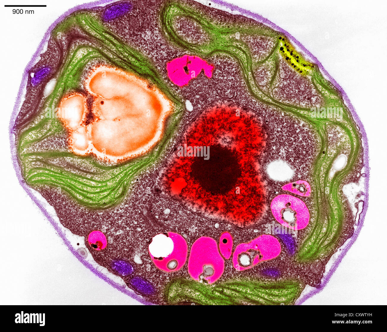
Plant Cell Electron Microscope Images / Plant Stem Section Under The Microscope Detail Microscopic Photography Things Under A Microscope Electron Microscope / As the wavelength of an electron can be up to 100,000 times shorter than that of visible light photons, electron microscopes have a higher resolving power than light microscopes and can reveal the structure of smaller objects.. Labeled diagram of plant cell, created with biorender.com. The invention of the electron microscope in the late 1930s and its refinement over the next half century permitted vastly improved visualization of cell and tissue fine structure. Tems are costly, large, cumbersome instruments that require special housing and maintenance. A scanning electron microscope (sem) is a type of electron microscope that produces images of a sample by scanning the surface with a focused beam of electrons.the electrons interact with atoms in the sample, producing various signals that contain information about the surface topography and composition of the sample. The typical characteristics that define the plant cell include cellulose, hemicellulose and pectin, plastids which play a major role in photosynthesis and storage of starch, large vacuoles responsible for regulating the cell turgor pressure.
The invention of the electron microscope in the late 1930s and its refinement over the next half century permitted vastly improved visualization of cell and tissue fine structure. Electron beams have shorter wavelengths than photons. As the wavelength of an electron can be up to 100,000 times shorter than that of visible light photons, electron microscopes have a higher resolving power than light microscopes and can reveal the structure of smaller objects. They require high voltages to increase the acceleration speed of electrons, which, once they pass through the sample (transmission), increase the. Jul 22, 2021 · figure:
Plant Cell Micrograph High Resolution Stock Photography And Images Alamy from c8.alamy.com A transmission electron microscope produces images via the interaction of electrons with a sample. (resolution is the degree of sharpness of an image.) figure 2 compares the magnification of a light microscope to that of a tem. A scanning electron microscope (sem) is a type of electron microscope that produces images of a sample by scanning the surface with a focused beam of electrons.the electrons interact with atoms in the sample, producing various signals that contain information about the surface topography and composition of the sample. A scanning electron microscope is a type of electron microscope that produces images of a sample by scanning it with a focused beam of electrons. Electron beams have shorter wavelengths than photons. An electron microscope is a microscope that uses a beam of accelerated electrons as a source of illumination. Mar 03, 2020 · with the advanced microscopes of today, such as the scanning electron microscope and transmission electron microscope, cell biologists are able to obtain detailed images of the smallest of cell structures and organelles. The organelles unique for plant cells are vacuole, cell wall, and chloroplast (shown in orange text).
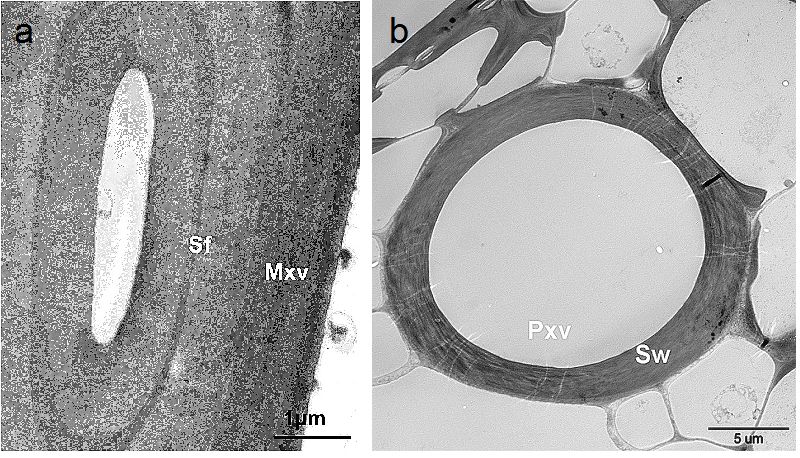
The transmission electron microscope (tem), the first type of em, has many commonalities with the optical microscope and is a powerful microscope, capable of producing images 1 nanometer in size.
The invention of the electron microscope in the late 1930s and its refinement over the next half century permitted vastly improved visualization of cell and tissue fine structure. As the wavelength of an electron can be up to 100,000 times shorter than that of visible light photons, electron microscopes have a higher resolving power than light microscopes and can reveal the structure of smaller objects. Labeled diagram of plant cell, created with biorender.com. The typical characteristics that define the plant cell include cellulose, hemicellulose and pectin, plastids which play a major role in photosynthesis and storage of starch, large vacuoles responsible for regulating the cell turgor pressure. A transmission electron microscope produces images via the interaction of electrons with a sample. They require high voltages to increase the acceleration speed of electrons, which, once they pass through the sample (transmission), increase the. in this figure the cell anatomy of animal and plant cells. An electron microscope is a microscope that uses a beam of accelerated electrons as a source of illumination. Advances in cell and molecular diagnostics , 2018 Jul 22, 2021 · figure: Electron microscopes, on the other hand, can produce much more highly magnified images because the beam of electrons has a smaller wavelength which creates images of higher resolution. As the wavelength of an electron can be up to 100,000 times shorter than that of visible light photons, electron microscopes have a higher resolving power than light microscopes and can reveal the structure of smaller objects. The shorter the wavelength of the illumination, the better the resolution.
An electron microscope is a microscope that uses a beam of accelerated electrons as a source of illumination. The organelles unique for plant cells are vacuole, cell wall, and chloroplast (shown in orange text). An electron microscope is a microscope that uses a beam of accelerated electrons as a source of illumination. (resolution is the degree of sharpness of an image.) figure 2 compares the magnification of a light microscope to that of a tem. Mar 03, 2020 · with the advanced microscopes of today, such as the scanning electron microscope and transmission electron microscope, cell biologists are able to obtain detailed images of the smallest of cell structures and organelles.
Scanning Electron Microscopy Of Roots Of Rice Oryza Sativa Plants Download Scientific Diagram from www.researchgate.net An electron microscope is a microscope that uses a beam of accelerated electrons as a source of illumination. A scanning electron microscope (sem) is a type of electron microscope that produces images of a sample by scanning the surface with a focused beam of electrons.the electrons interact with atoms in the sample, producing various signals that contain information about the surface topography and composition of the sample. in this figure the cell anatomy of animal and plant cells. The animal cell and plant cell share many organelles in common, such as a nucleus, er, cytosol, lysosomes, golgi apparatus, cell membrane, and ribosomes. They require high voltages to increase the acceleration speed of electrons, which, once they pass through the sample (transmission), increase the. A transmission electron microscope produces images via the interaction of electrons with a sample. Mar 03, 2020 · with the advanced microscopes of today, such as the scanning electron microscope and transmission electron microscope, cell biologists are able to obtain detailed images of the smallest of cell structures and organelles. The organelles unique for plant cells are vacuole, cell wall, and chloroplast (shown in orange text).
The organelles unique for plant cells are vacuole, cell wall, and chloroplast (shown in orange text).
Advances in cell and molecular diagnostics , 2018 Labeled diagram of plant cell, created with biorender.com. Mar 03, 2020 · with the advanced microscopes of today, such as the scanning electron microscope and transmission electron microscope, cell biologists are able to obtain detailed images of the smallest of cell structures and organelles. As the wavelength of an electron can be up to 100,000 times shorter than that of visible light photons, electron microscopes have a higher resolving power than light microscopes and can reveal the structure of smaller objects. in this figure the cell anatomy of animal and plant cells. The organelles unique for plant cells are vacuole, cell wall, and chloroplast (shown in orange text). The typical characteristics that define the plant cell include cellulose, hemicellulose and pectin, plastids which play a major role in photosynthesis and storage of starch, large vacuoles responsible for regulating the cell turgor pressure. Tems are costly, large, cumbersome instruments that require special housing and maintenance. As the wavelength of an electron can be up to 100,000 times shorter than that of visible light photons, electron microscopes have a higher resolving power than light microscopes and can reveal the structure of smaller objects. The animal cell and plant cell share many organelles in common, such as a nucleus, er, cytosol, lysosomes, golgi apparatus, cell membrane, and ribosomes. They require high voltages to increase the acceleration speed of electrons, which, once they pass through the sample (transmission), increase the. The shorter the wavelength of the illumination, the better the resolution. Electron beams have shorter wavelengths than photons.
The animal cell and plant cell share many organelles in common, such as a nucleus, er, cytosol, lysosomes, golgi apparatus, cell membrane, and ribosomes. Mar 03, 2020 · with the advanced microscopes of today, such as the scanning electron microscope and transmission electron microscope, cell biologists are able to obtain detailed images of the smallest of cell structures and organelles. The invention of the electron microscope in the late 1930s and its refinement over the next half century permitted vastly improved visualization of cell and tissue fine structure. An electron microscope is a microscope that uses a beam of accelerated electrons as a source of illumination. in this figure the cell anatomy of animal and plant cells.
Ultrastructure And Topochemistry Of Plant Cell Wall By Transmission Electron Microscopy Intechopen from www.intechopen.com They require high voltages to increase the acceleration speed of electrons, which, once they pass through the sample (transmission), increase the. The organelles unique for plant cells are vacuole, cell wall, and chloroplast (shown in orange text). The transmission electron microscope (tem), the first type of em, has many commonalities with the optical microscope and is a powerful microscope, capable of producing images 1 nanometer in size. (resolution is the degree of sharpness of an image.) figure 2 compares the magnification of a light microscope to that of a tem. Tems are costly, large, cumbersome instruments that require special housing and maintenance. The invention of the electron microscope in the late 1930s and its refinement over the next half century permitted vastly improved visualization of cell and tissue fine structure. Labeled diagram of plant cell, created with biorender.com. Electron microscopes, on the other hand, can produce much more highly magnified images because the beam of electrons has a smaller wavelength which creates images of higher resolution.
The organelles unique for plant cells are vacuole, cell wall, and chloroplast (shown in orange text).
As the wavelength of an electron can be up to 100,000 times shorter than that of visible light photons, electron microscopes have a higher resolving power than light microscopes and can reveal the structure of smaller objects. As the wavelength of an electron can be up to 100,000 times shorter than that of visible light photons, electron microscopes have a higher resolving power than light microscopes and can reveal the structure of smaller objects. (resolution is the degree of sharpness of an image.) figure 2 compares the magnification of a light microscope to that of a tem. The animal cell and plant cell share many organelles in common, such as a nucleus, er, cytosol, lysosomes, golgi apparatus, cell membrane, and ribosomes. A transmission electron microscope produces images via the interaction of electrons with a sample. The invention of the electron microscope in the late 1930s and its refinement over the next half century permitted vastly improved visualization of cell and tissue fine structure. Electron microscopes, on the other hand, can produce much more highly magnified images because the beam of electrons has a smaller wavelength which creates images of higher resolution. An electron microscope is a microscope that uses a beam of accelerated electrons as a source of illumination. Jul 22, 2021 · figure: An electron microscope is a microscope that uses a beam of accelerated electrons as a source of illumination. Tems are costly, large, cumbersome instruments that require special housing and maintenance. The organelles unique for plant cells are vacuole, cell wall, and chloroplast (shown in orange text). Labeled diagram of plant cell, created with biorender.com.
Share :
Post a Comment
for "Plant Cell Electron Microscope Images / Plant Stem Section Under The Microscope Detail Microscopic Photography Things Under A Microscope Electron Microscope / As the wavelength of an electron can be up to 100,000 times shorter than that of visible light photons, electron microscopes have a higher resolving power than light microscopes and can reveal the structure of smaller objects."


Post a Comment for "Plant Cell Electron Microscope Images / Plant Stem Section Under The Microscope Detail Microscopic Photography Things Under A Microscope Electron Microscope / As the wavelength of an electron can be up to 100,000 times shorter than that of visible light photons, electron microscopes have a higher resolving power than light microscopes and can reveal the structure of smaller objects."